The Eyes as a Window to Alzheimer’s: New Research Offers Hope for Early Detection
Learn how scientific advances mean eye exams could be a potential tool for early Alzheimer’s diagnosis.

When we think about Alzheimer’s disease, we often associate it with memory loss and cognitive decline. However, new research suggests that the eyes may provide an early warning system for detecting Alzheimer’s long before symptoms appear.
Scientists have discovered that proteins linked to the disease, such as amyloid, can be detected in the eyes, opening the door for potential early diagnosis through retinal exams.
Aging and visual perception
Visual perception begins when light enters the eyes and transforms into signals that reach the brain, where they’re processed and consciously experienced. The eyes constantly gather environmental data, and the brain interprets it, shaping how we interact with the world. Aging, however, alters both our eyes and brains.
Conditions like glaucoma, cataracts, macular degeneration, diabetes, or age-related changes in parts of the eye, such as the retina or macula, disrupt the flow of visual information to the brain. This distorted information can contribute to cognitive issues like dementia. Conversely, brain changes from dementia or Alzheimer’s disease can scramble how we process visual data, distorting our perception and understanding of the world.
How Alzheimer’s disease can affect vision
As we age, our ability to process visual information declines. Medical conditions can worsen these issues, leading to symptoms such as:
- Blurred vision
- Slower adjustment to light
- Reduced peripheral vision
- Difficulty processing distance and three-dimensional objects
Visual perception changes in Alzheimer’s disease and dementia are often linked to structural alterations in the brain. These changes impact several areas critical for visual processing, including the occipital lobe, responsible for interpreting visual stimuli, and the parietal lobe, responsible for spatial awareness and coordination.
Some common visual-perceptual difficulties caused by these structural changes include:
- Decreased sensitivity to contrast, especially in low-light or cluttered environments
- Difficulty recognizing certain colors
- Problems with gaze direction
- Reduced depth perception
Posterior cortical atrophy, also known as Benson’s syndrome, is a degenerative disease that damages the brain’s visual center. For a majority of people, the cause of posterior cortical atrophy is Alzheimer’s disease. It is characterized by visual distortions and hallucinations that may include:
- Difficulty recognizing objects, faces, and colors
- Distortions, sensitivity to light, and visual hallucinations
- Trouble judging distances and reaching for objects
- Difficulty reading a line of text
- Difficulty writing, spelling, and performing calculations
These visual-perceptual changes can contribute to anxiety, confusion, and unusual behaviors in people with Alzheimer’s, leaving loved ones and caregivers puzzled and concerned. Understanding how Alzheimer’s impacts vision is crucial in providing better care and creating a supportive environment for individuals affected by the disease.
My previous blog post on dementia and eyesight digs deeper into these visual changes and the behaviors that can result from them. I also offer tips for those caring for a loved one with both dementia and visual perception changes.
The connection between retinal changes and brain health
The retina is a layer of cells that lines the back of your eye. It plays a crucial role in vision, converting light into signals sent to the brain.
The retina is an extension of the brain. The brain and retina share many characteristics, including susceptibility to neurodegenerative conditions. These similarities make the eyes valuable windows into brain health. Researchers have found that amyloid plaques, a hallmark of Alzheimer’s, can accumulate in the retina in the same way they do in the brain.
A recent study examined the retina and brain tissues of individuals with mild cognitive impairment and Alzheimer’s disease. Researchers found significant increases in amyloid beta-protein and novel intraneuronal amyloid-beta oligomers — small protein clusters that form inside nerve cells.
The research found that these proteins were closely associated with tissue damage and retinal inflammation. These eye changes matched the severity of brain damage and cognitive decline, suggesting that they could help detect Alzheimer’s disease.
Retinal biomarkers of Alzheimer’s disease

Emerging research has highlighted the potential of retinal biomarkers in identifying Alzheimer’s disease in its early stages. Biomarkers are measurable warning signs for a disease. Retinal imaging technology such as optical coherence tomography, a non-invasive test that uses light waves to take photos of your retina, plus other computational advances, has helped propel research forward.
Researchers have discovered multiple structural changes in the retinas of those with Alzheimer’s disease:
- Thinning of the retinal nerve fiber layer: The retinal nerve fiber layer, or the thin layer of nerve fibers that covers the retina, particularly around the eye’s optic disc and macula, is thinner in people with cognitive decline.
- Degeneration of ganglion cell layer: Some patients with Alzheimer’s disease have lost retinal ganglion cells, which transmit visual information from the retina to the brain.
- Changes in retinal blood vessels: Retinal blood flow and vessel density changes have been linked to Alzheimer’s, suggesting that neurovascular dysfunction contributes to the disease.
- Amyloid and tau deposits in the retina: Some studies indicate that amyloid plaques can be detected in retinal tissues.
Additionally, a new study found that the presence of the APOE4 gene, which is a strong genetic risk factor for Alzheimer’s disease in humans, is associated with retinal degeneration in mice. The findings suggested that changes associated with Alzheimer’s disease manifest in the eyes before symptoms appear, underscoring the potential of the retina as a biomarker for early detection.
These retinal changes may serve as valuable non-invasive biomarkers for early Alzheimer’s detection, providing an alternative to costly and invasive brain imaging techniques.
How changes in eyesight and pupil response can offer clues about brain health
Studies have shown that reduced contrast sensitivity, or the ability to see fine details, is associated with amyloid and tau deposits in the brain. A simple, non-invasive vision test called frequency doubling technology can help measure contrast sensitivity. Scientists are exploring whether this test could be used as a biomarker for Alzheimer’s disease since vision changes like these may appear before memory problems or other symptoms of dementia begin.
Changes in how our pupils get bigger or smaller — called pupillary responses — can give us clues about brain health. In people with Alzheimer’s disease or mild cognitive impairment, which often comes before Alzheimer’s, these responses can look different. Studies have found that people with mild cognitive impairment often have larger pupil dilation when remembering and thinking.
These findings might mean the brains of individuals with mild cognitive impairment are working harder to keep up, even if they don’t show obvious memory issues yet. These pupil changes can also help distinguish between people with mild cognitive impairment and those with typical brain function. Researchers believe these changes may be linked to activity in a part of the brain called the locus coeruleus, one of the first areas affected by Alzheimer’s disease.
The promise of retinal exams for early Alzheimer’s detection
While more studies are needed to validate these findings, the potential of eye exams in Alzheimer’s detection is clear. The discovery of biomarkers paves the way for future diagnostic tools that could make early detection more straightforward and widespread.
Advances in retinal imaging have already shown that amyloid deposits and other markers linked to cognitive impairment can be found in patients’ eyes. As research progresses, retinal imaging holds the potential to be validated and combined with other key markers, such as cognitive assessments and brain imaging techniques, to better identify individuals at risk. This integration could lead to earlier interventions and improved outcomes for patients and their families.
Early Alzheimer’s detection empowers patients and loved ones
Early detection is key to better management of Alzheimer’s disease. Recent progress in biomarker research presents exciting opportunities for early detection and screening, which could allow for interventions when treatments to modify the disease may have the greatest impact.
This also underscores the importance of regular eye exams — not just for vision health but as a proactive measure against neurodegenerative diseases. Studies suggest that retinal changes could even be detected in individuals with mild cognitive impairment long before significant memory loss occurs, reinforcing the value of eye exams as an accessible diagnostic tool.
Keeping an eye on your vision might just mean keeping an eye on your brain health, too. With ongoing scientific advancements, the future of Alzheimer’s detection could be as simple as looking into your eyes.
Comprehensive outpatient care for all stages of memory loss
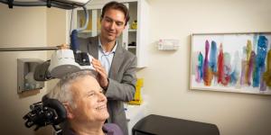
At the Deanna and Sidney Wolk Center for Memory Health, we offer eye exams as part of a routine evaluation of brain health, memory problems, and dementia. If you’re concerned about symptoms of cognitive decline in yourself or a loved one, we are here for you.
We offer comprehensive diagnosis and assessment services and can help you put together an individualized plan and resources to maximize the safety and well-being of you or your loved one. We also offer extensive support services for caregivers and families of people with dementia. Interested in learning more? Call us at 617-363-8600 or contact us today.
Join Our Community: Subscribe to the Hebrew SeniorLife blog for weekly insights on healthy aging and senior living.
Blog Topics
Learn More
Wolk Center for Memory Health
The Deanna and Sidney Wolk Center for Memory Health at Hebrew SeniorLife provides outpatient memory care services, in person and virtually, for people living with cognitive symptoms — and for their families and caregivers.
Memory Care
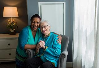
Our care team helps people with advanced Alzheimer's disease and other forms of dementia feel safe and stimulated, while maintaining a connection to community.